Lab 8 -- Secondary xylem
These photos were taken from sections made by students in the lab
on secondary xylem, where we looked at woody stems of young pine and
several dicot flowering plants. The woodiness of the tissue made
it harder to get thin sections, but many of them showed the various
cell types quite nicely.
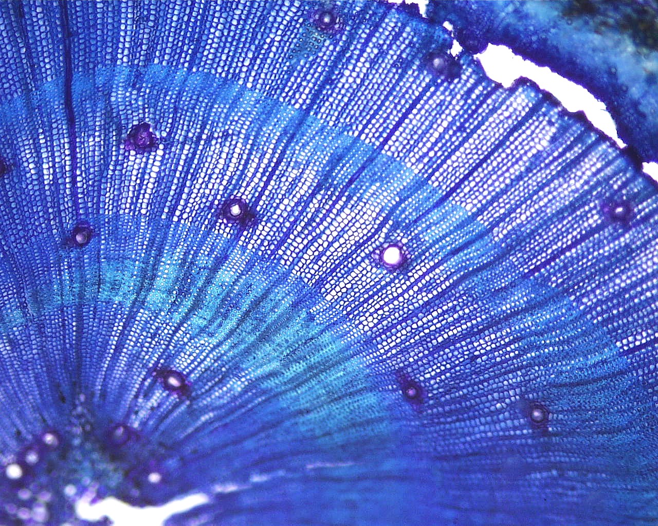
In this first set of images, we see transverse sections of young pine
stems. Most of the stem cross-section was made up of secondary
xylem, containing almost exclusively tracheids. The first image
below shows three growth rings of secondary xylem. Notice how the
tracheids have thinner walls early in the growth ring (towards the
center of the stem) and the walls are thicker in the later part of the
growth ring.
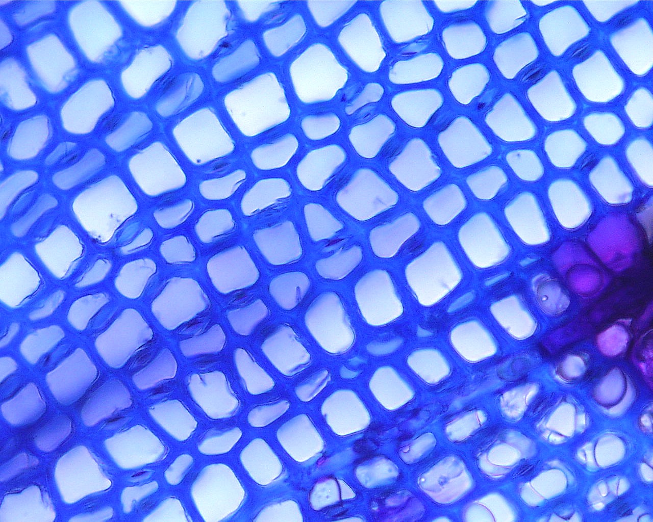
Here is a single growth ring of the pine. The changes in tracheid
wall thickness described above are more apparent. Also notice how
the cells get narrower (in the radial direction) in the latewood part
of the growth ring (towards the right). The rays are
uniseriate. Notice the resin duct near the top center.

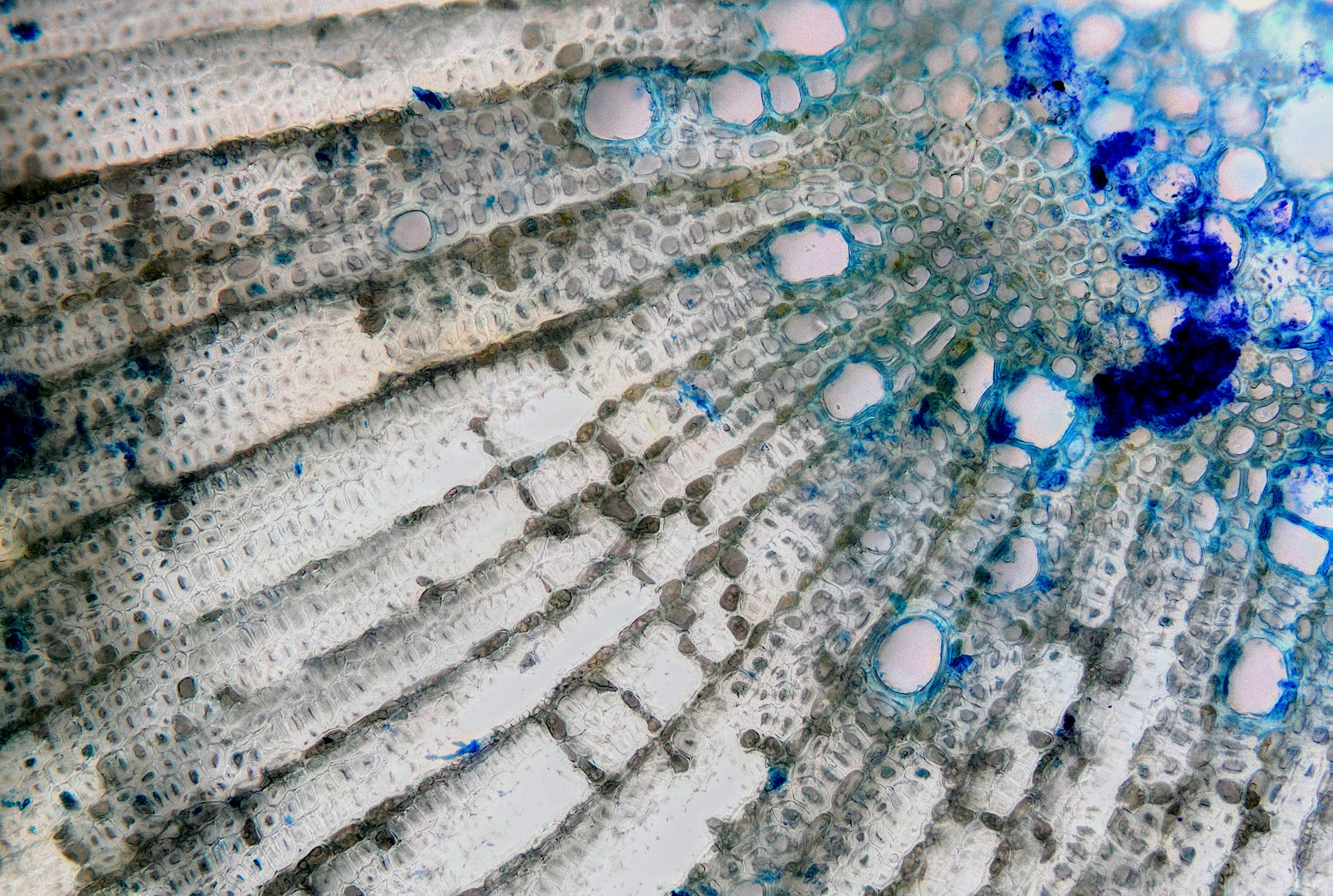
Here's a closer view of the tracheids in pine secondary xylem.
Notice how the pits are located exclusively on the radial walls (the
ray at the lower right should tell you which way the tissue is
oriented). There is a tracheid near the center where the torus in
the pit membrane is visible as a thin dark disk.
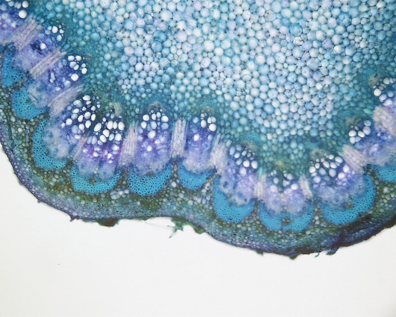
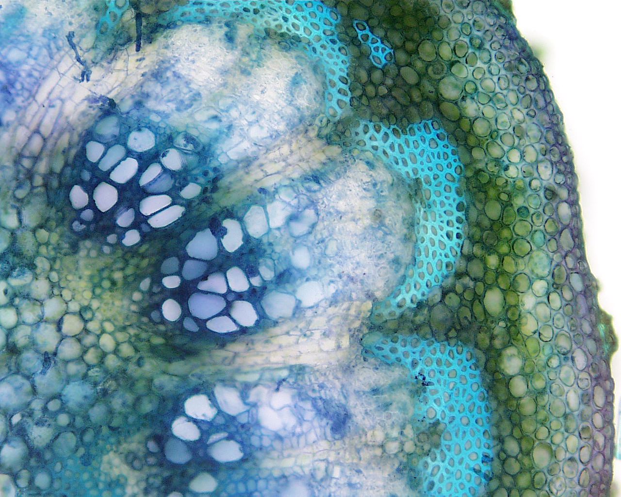
These are images from young sycamore stems. You can still see
parts of the old vascular bundles where a vascular cambium developed
and then began producing secondary xylem and phloem. Note the
wide rays of these secondary tissues.

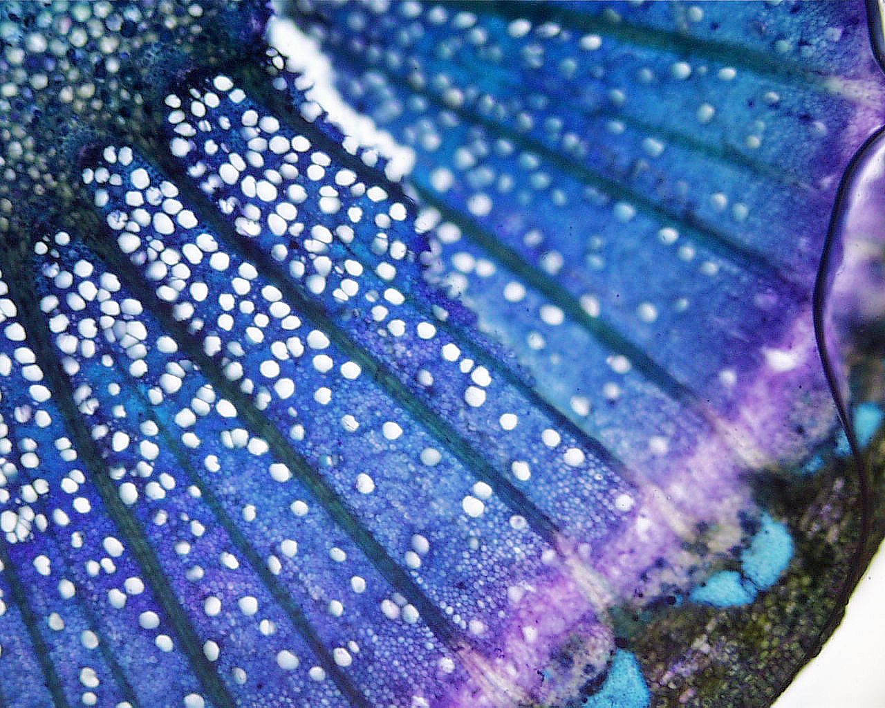
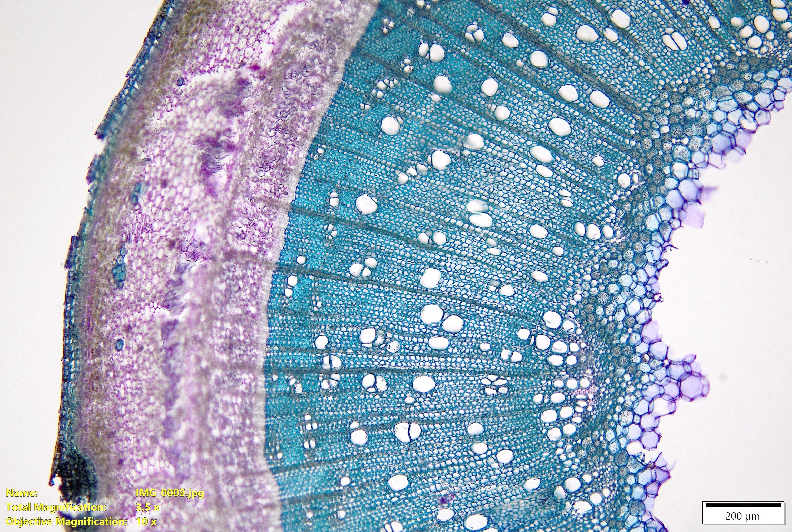
This is an image from a one-year-old sycamore stem (one growth
ring). The xylem appears to be diffuse porous - vessel diameter
is roughly constant across the growth ring and they are distributed
evenly. Notice the fairly wide rays.
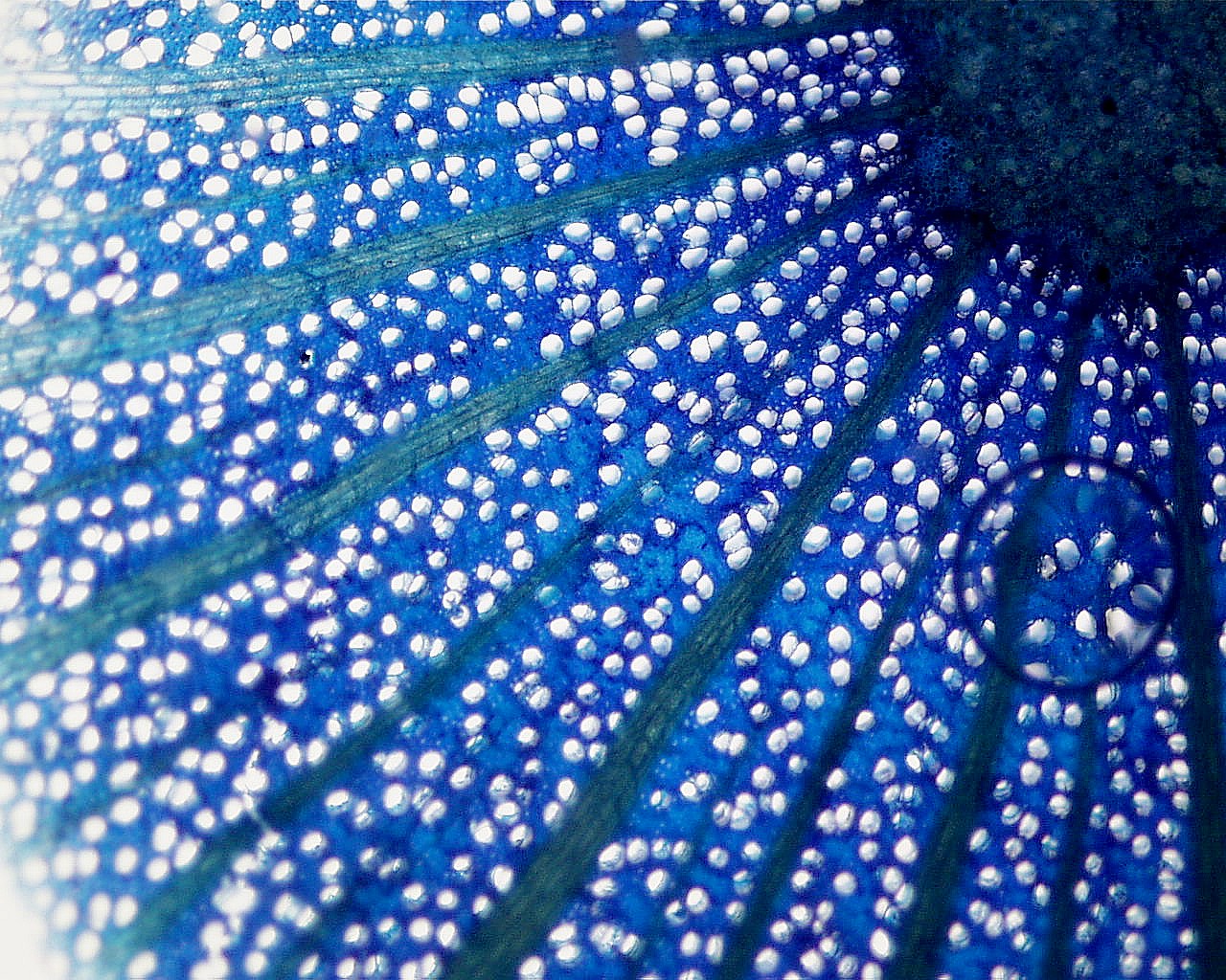
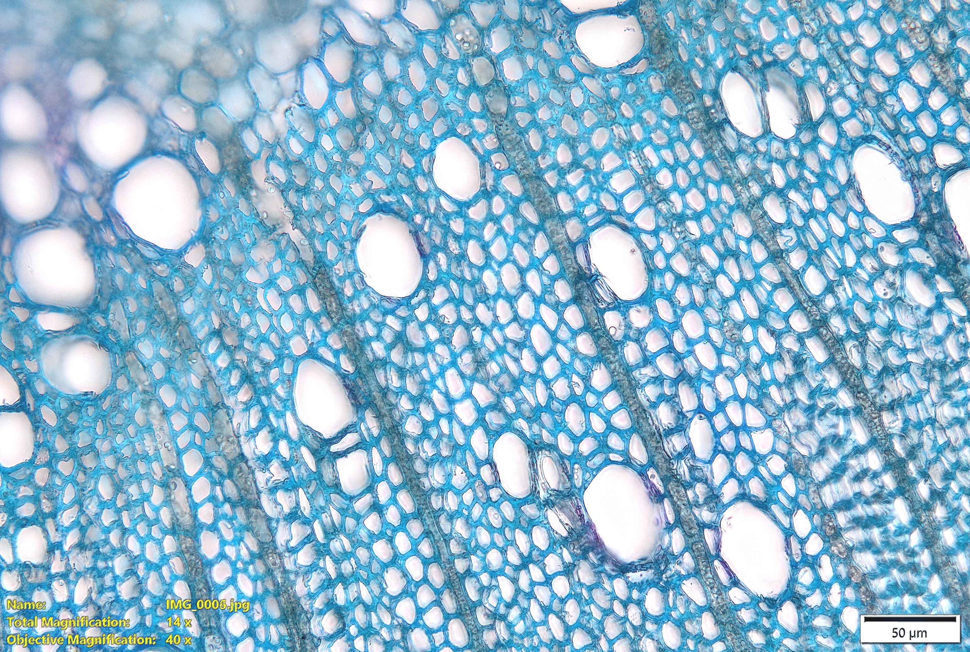
In this closer view of sycamore xylem, you can see the multiseriate
rays (lots of starch). Most of the cells around the vessels have
fairly thick walls and might be fibers, although a longitudinal section
would be useful for checking the wall pitting.
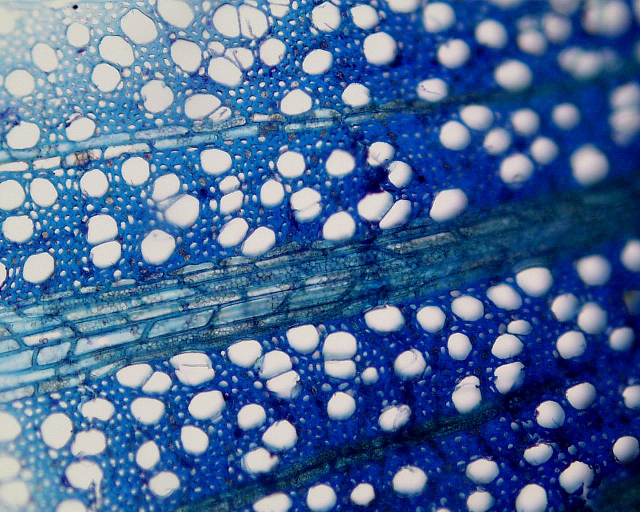
The next image is a transverse view of secondary xylem from a live oak stem (Quercus virginiana). There are many tangential bands of xylem parenchyma present in the secondary xylem.
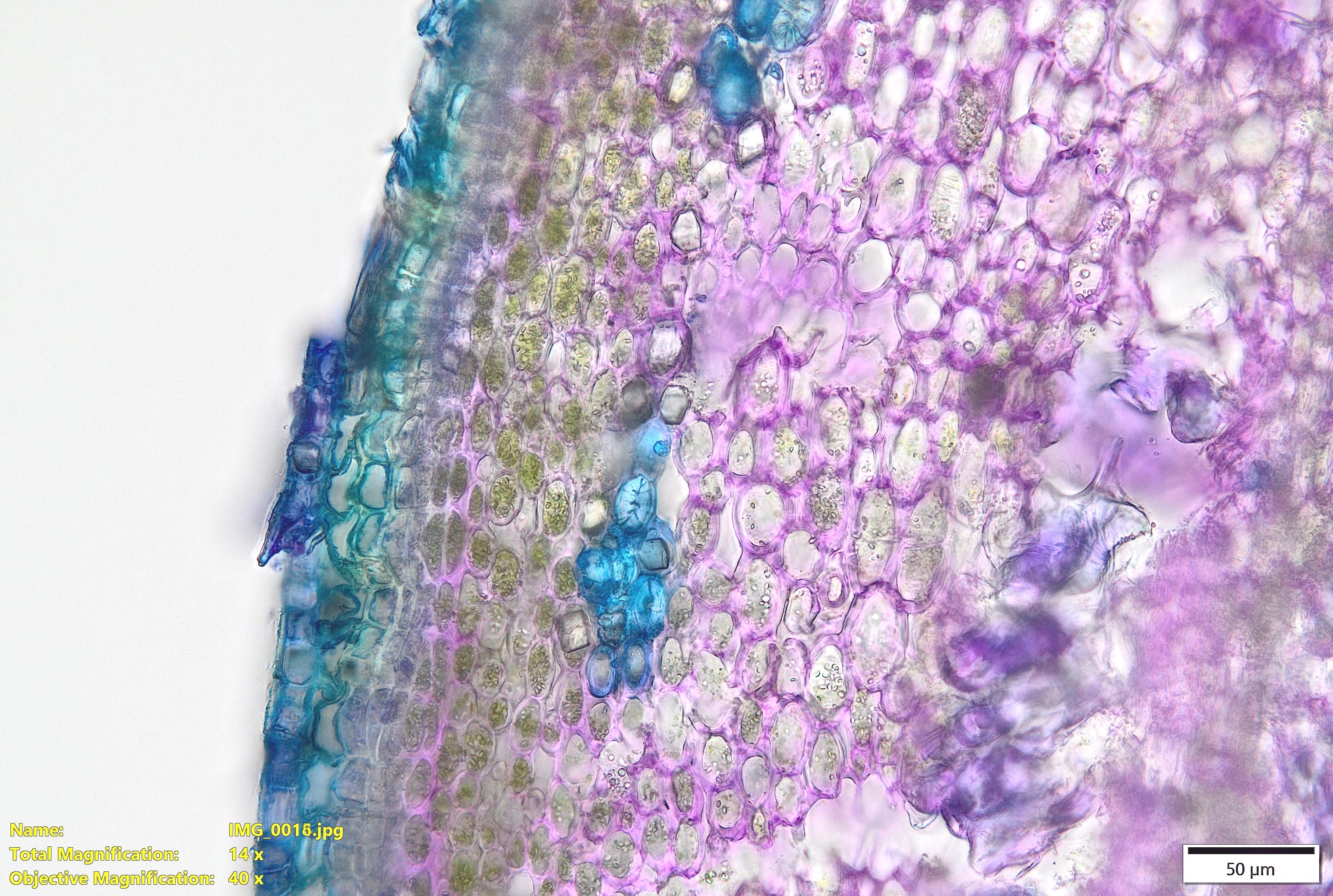
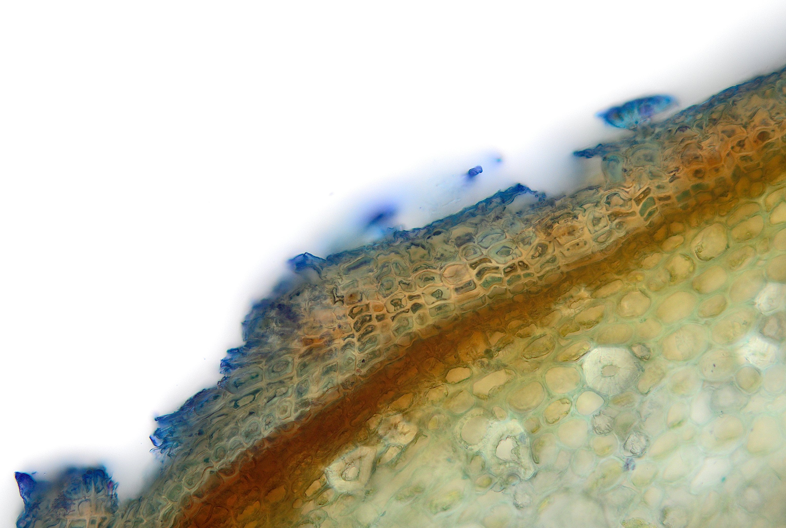
These last two live oak views show periderm at the edge of the stem.

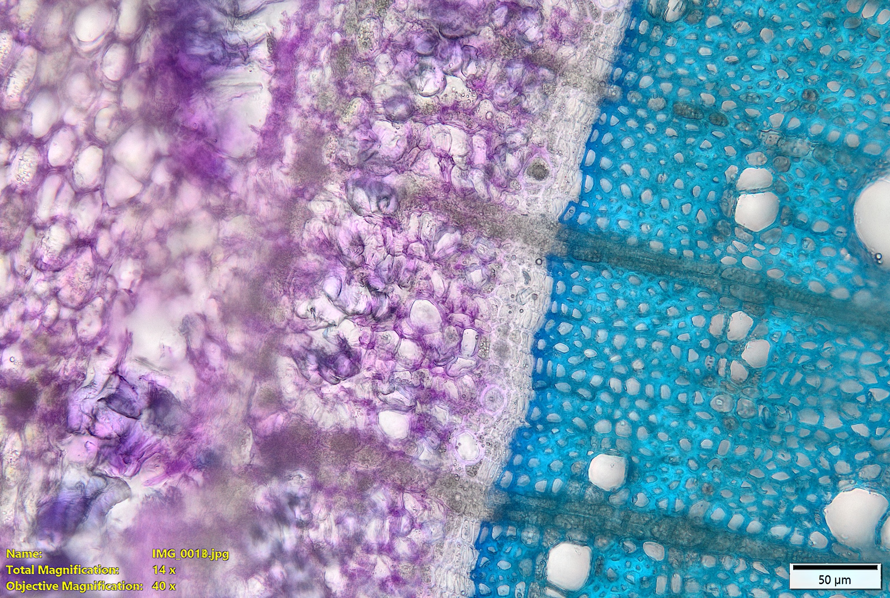
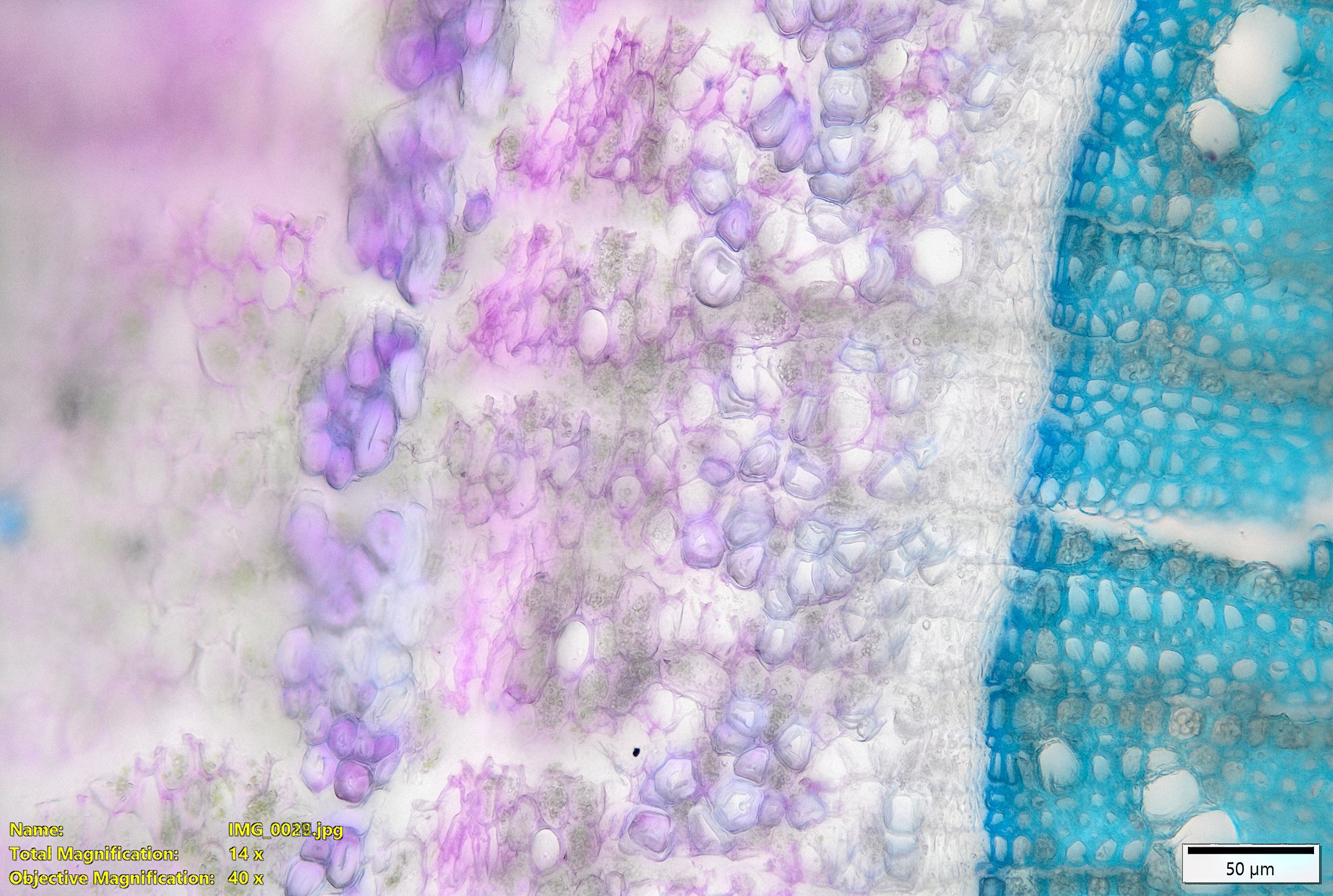
The next five images show transverse sections through a mulberry stem.
Notice all the starch grains inside the rays cells.
The next image shows the transition between secondary xylem and phloem.
Next is a view of the secondary phloem of the mulberry stem.
Lastly is a view of the edge of the stem with a photosynthetic cortex and the first periderm forming inside the epidermis.