Lab 3 -- Tissues

Here are the photos taken by students in class based on their hand sections from a variety of plant tissues.  Although some of the sections could always be thinner, many of the important characteristics of the epidermis, xylem, and phloem tissues can be seen.

Here's an epidermal peel from the purple onion.  Nice job peeling, all these cells are intact (they haven't lost the purple pigment).  Notice how the tissue does have stomata, but they are very few in number.  Also, this is a monocot with a fairly ordered arrangement of the cells (compare to the spinach epidermis from last week's images).


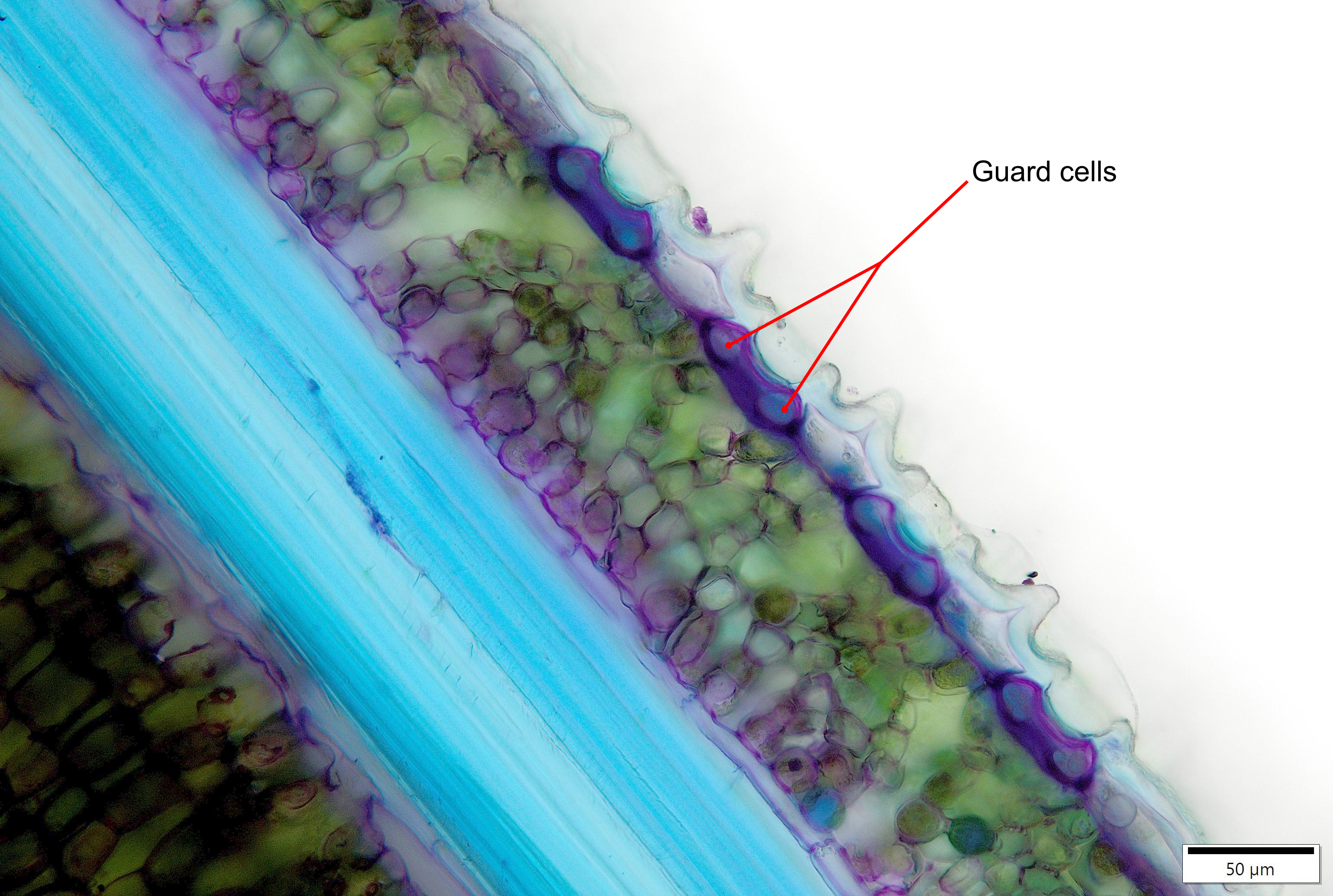
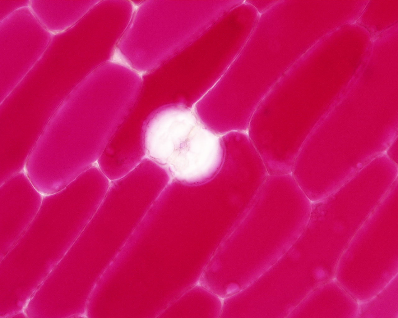
Here's a closeup of that stomata with a pair of guard cells, although they ended up a bit overexposed.


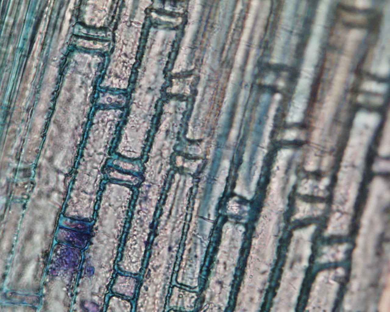
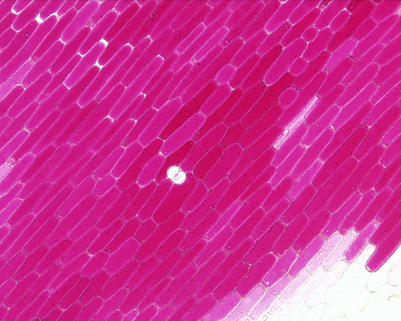
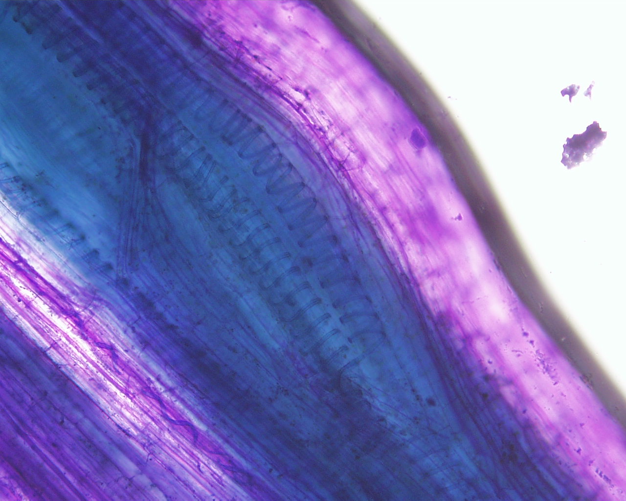
These next three images show the surface of the monocot leaf.  You can see the ordered arrangement of cells.  The guard cells are the small square-shaped cells at the ends of the brick-shaped other epidermal cells.


Here's an image of a commercially prepared slide from a pine stem tip.  It shows a longitudinal view with several xylem tracheids.  You can see the developmental progression (right-to-left) of tracheids having annular to helical (spiral) thickenings and then bordered pits.


These next two are "squashes" of celery petiole vascular bundles.  You can see the helical thickenings of the tracheary elements, although there are lots of other cells obstructing the view a bit.


These next images are sections through a squash stem (Cucurbita sp.) showing an unusual vascular bundle form where there is phloem tissue on both sides of the xylem - we'll come to stem structures in a few weeks for more about this...For now, the dark blue stained cells in the middle are xylem vessels and the adjacent small purple cells (above and below) are phloem.


Here's a closer view showing xylem and one side of the phloem: