Lab 6 -- Stems I
These photos were taken from sections made by students in the first lab
on stems, which looked at pea and corn as dicot and monocot examples.
The corn seedlings used in lab where a bit younger than would have been
ideal, but the students collected a range of sections from regions that were
mostly root-like through the root-stem transition, to sections made from the
stem (though a very young stem) to sections showing closely folded leaves
(beyond the shoot apical meristem).
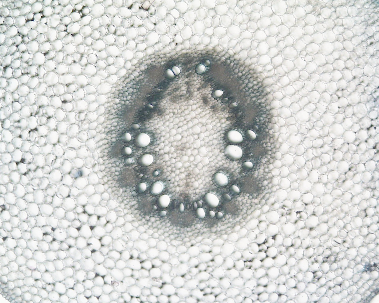
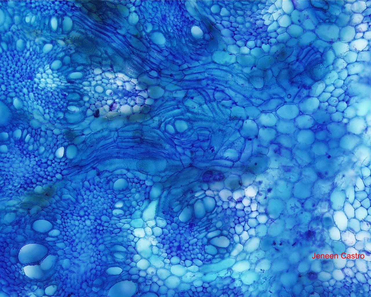
Here are a few images of corn that are probably closer to the kind of vascular
system we saw in corn roots - remember the polyarch arrangement? The
location of the wider metaxylem towards the center (exarch)?.

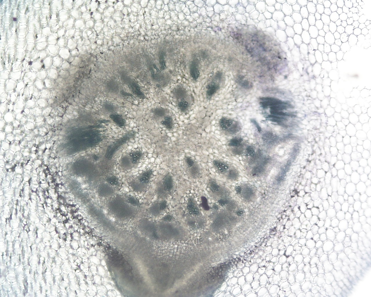
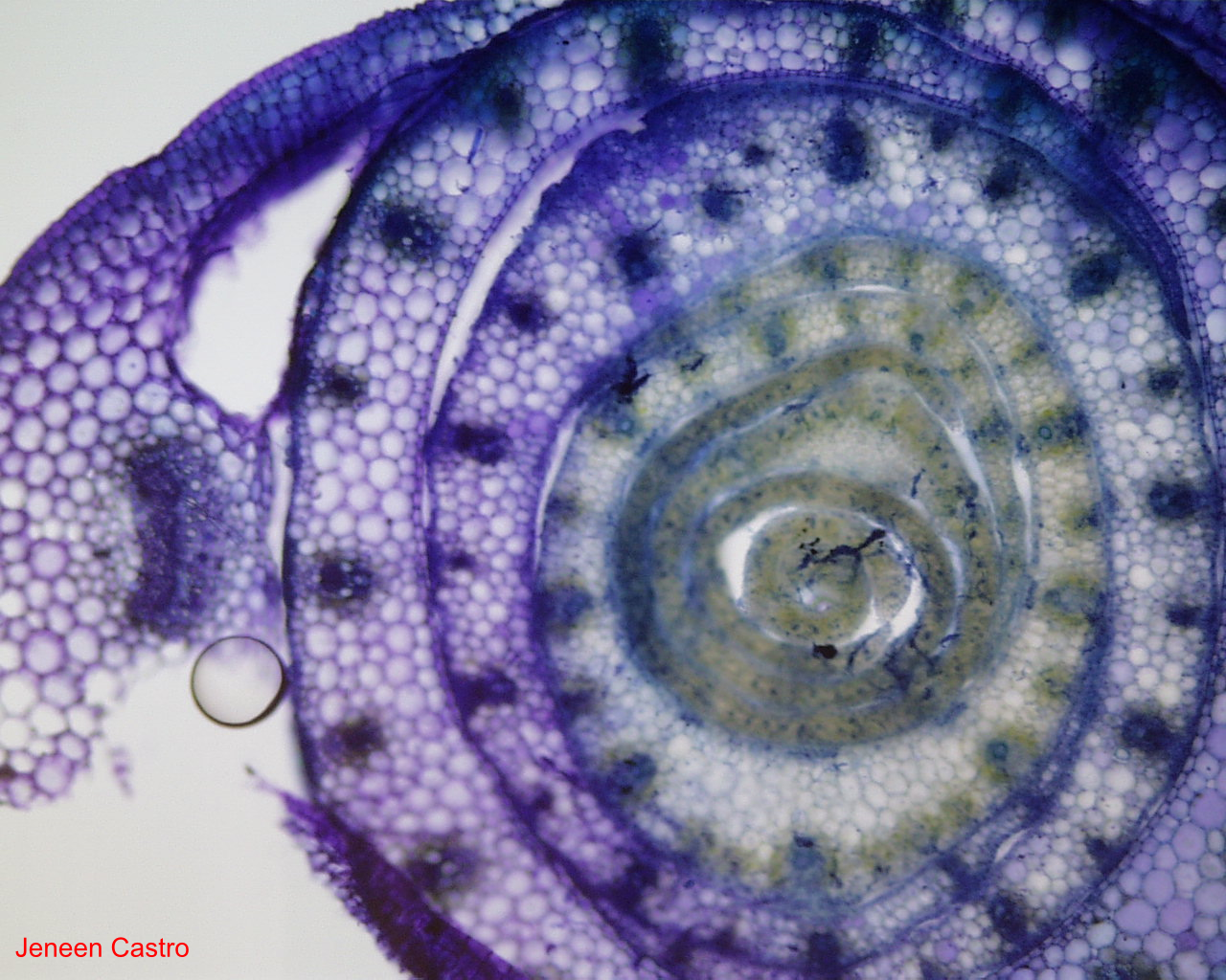
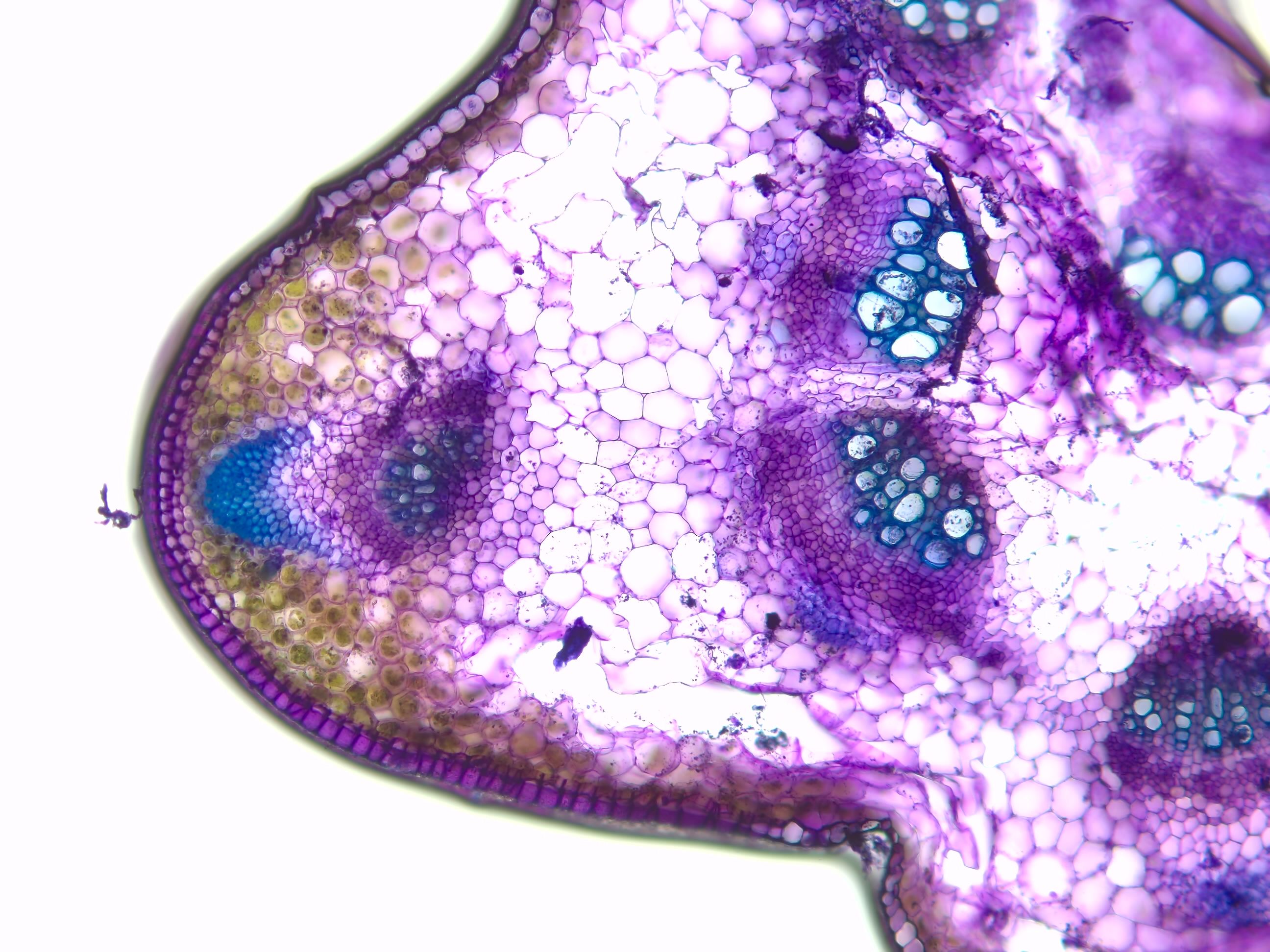
Here's an image from the root - stem transition. Now we see
discrete vascular bundles forming, but they are not quite like the "scattered"
bundle arrangement found in more mature corn stems (see lab handout image).
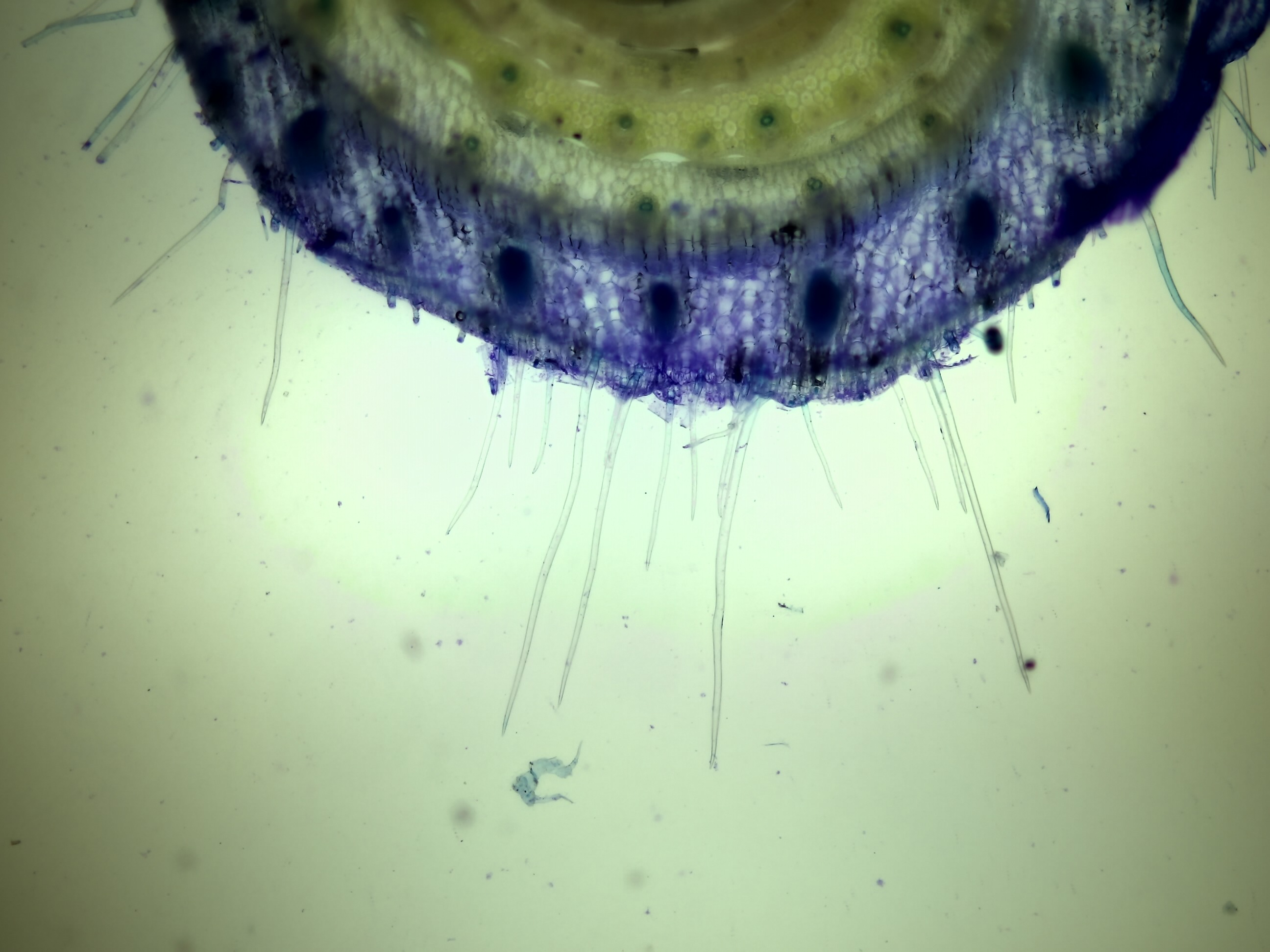
A couple of these images had what I am guessing might be an adventitious
root forming and pushing out through the cortex.
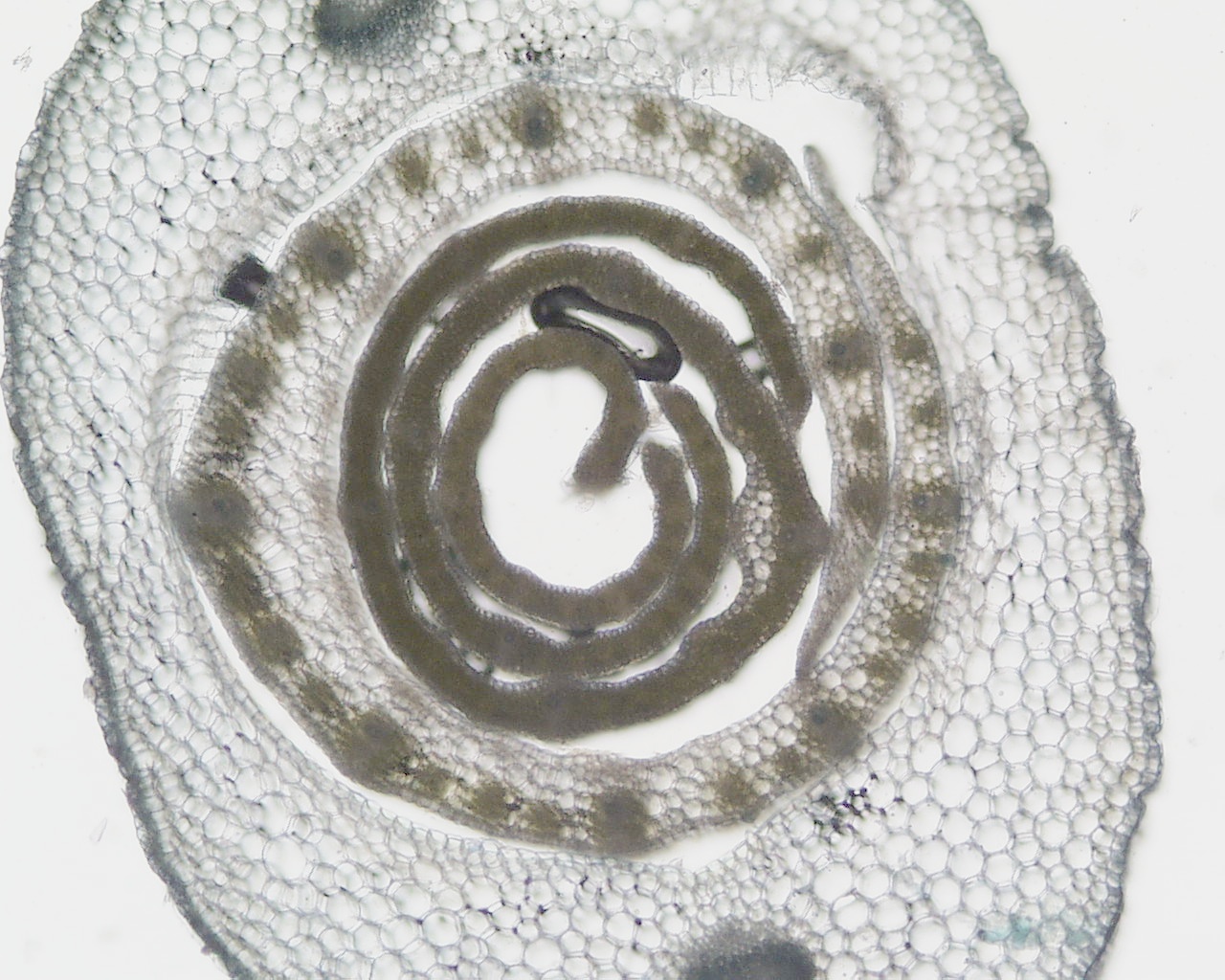
Beyond the actual stem apex, you would get sections that show corn leaves
rolled up together below where they spread out to form functional leaves.
You should be able to identify the vascular bundles in these leaves.
Think about how those individual vascular bundles in the stem below
must be connecting together with these bundles in the leaves.
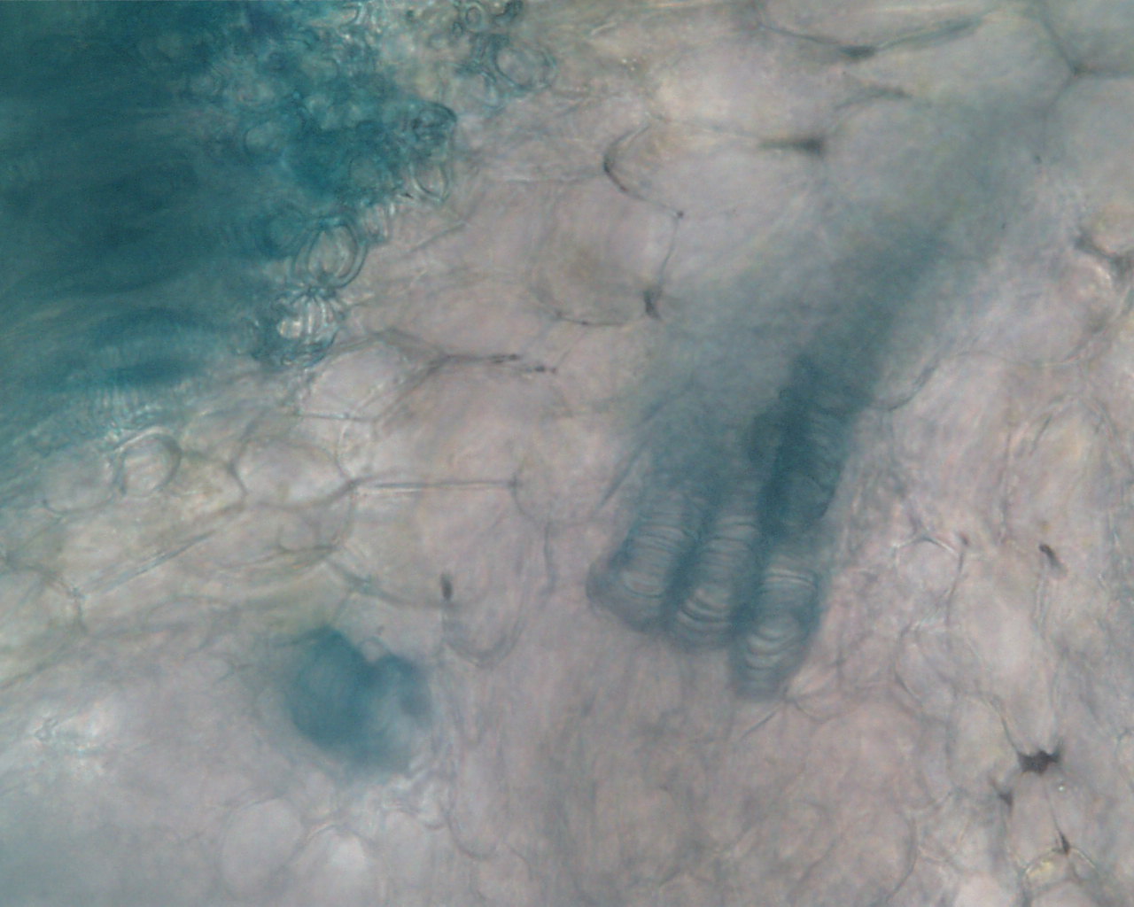
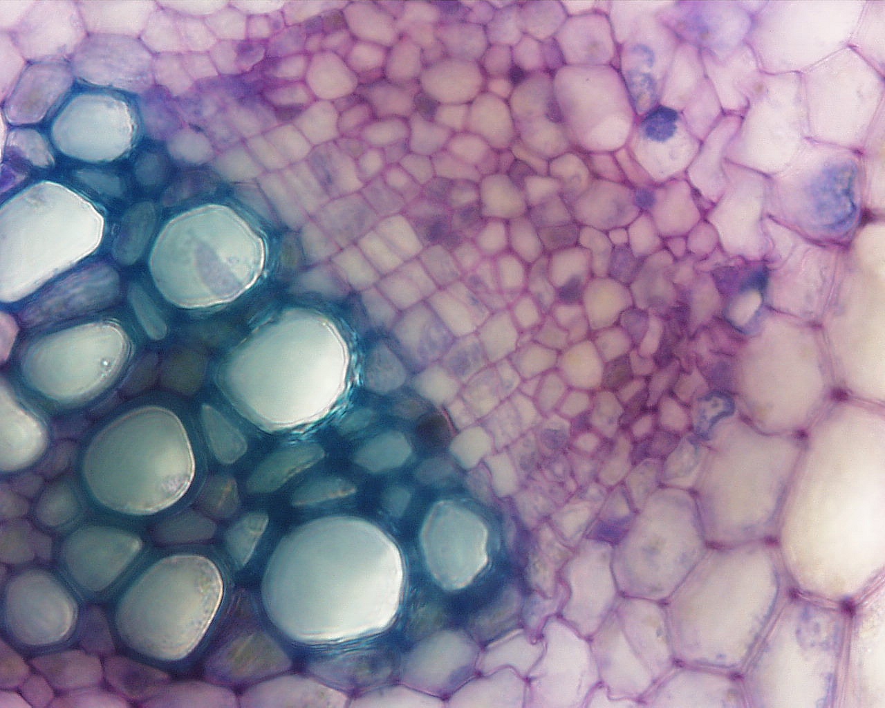
The following two images show close-ups of the vascular bundles in the
stem where they are turned enough to show the typical kind of secondary thickening
(helical) that one might find in protoxylem tracheary elements.

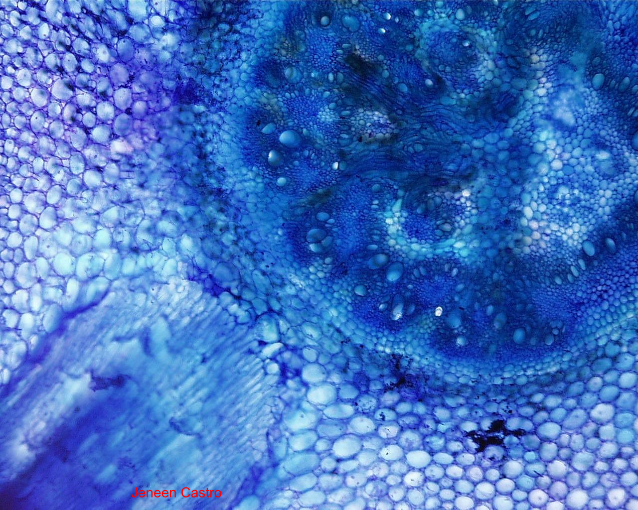
Now, moving on to the pea (dicot) seedlings. Here is a view of the
stem cross-section. There are several vascular bundles near the
center surrounding pith and surrounded by cortex. Many of these images
will also show other vascular bundles further out.
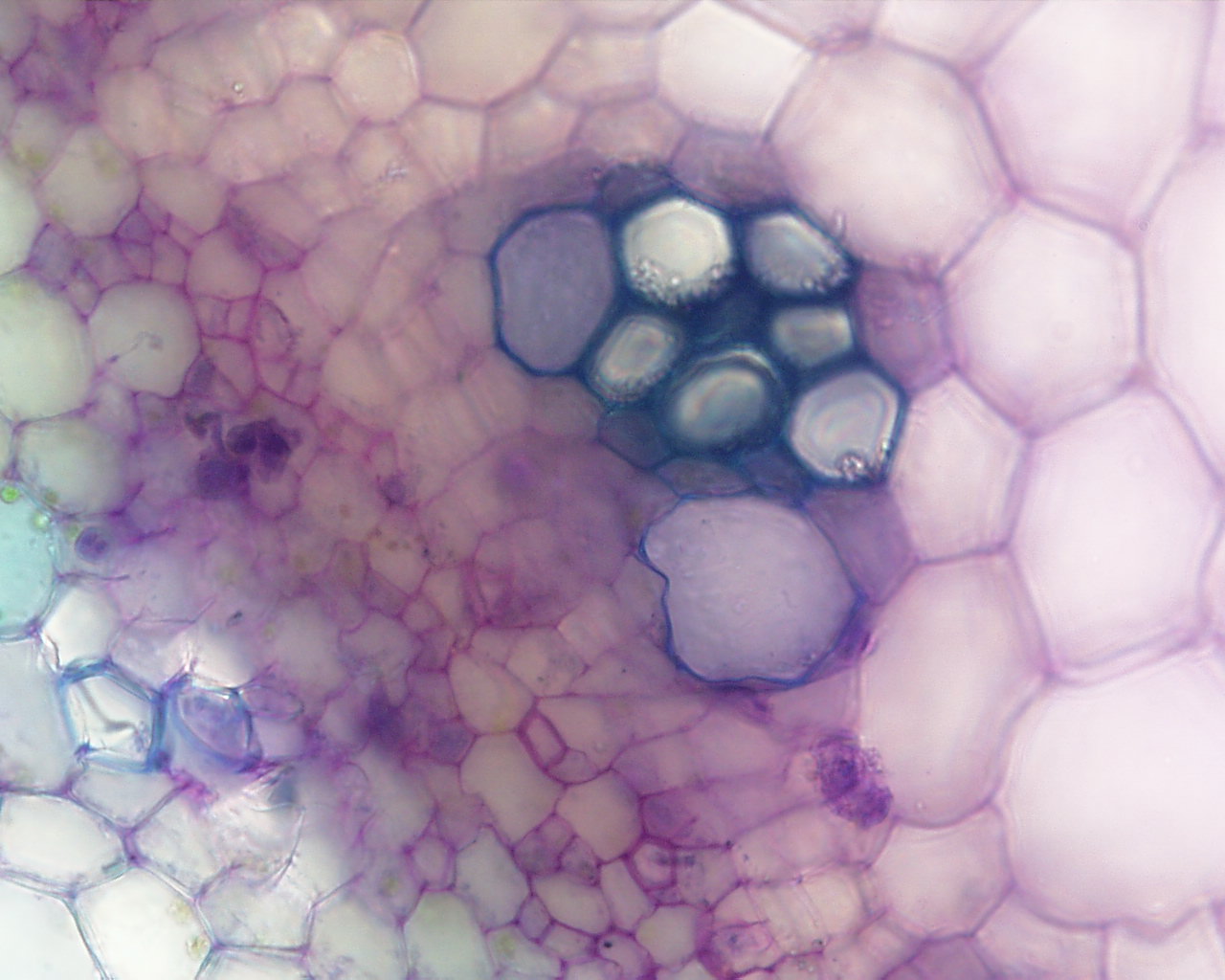
These next two images show details of individual vascular bundles. You
should be able to identify xylem and phloem. Notice that a cambium is
developing in the larger bundle.
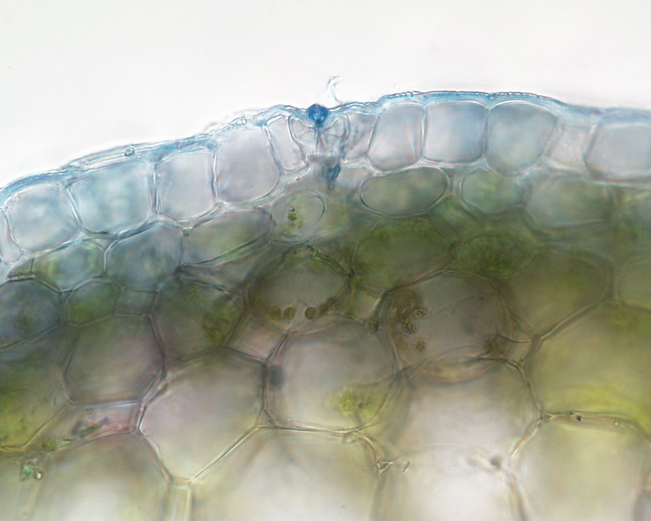
You should have also observed the outer parts of the stem, where the cortex
was photosynthetic (the outer 2 or 3 layers with chloroplasts) and the epidermis
was made from tightly connected cells. Look for the guard cell pairs.

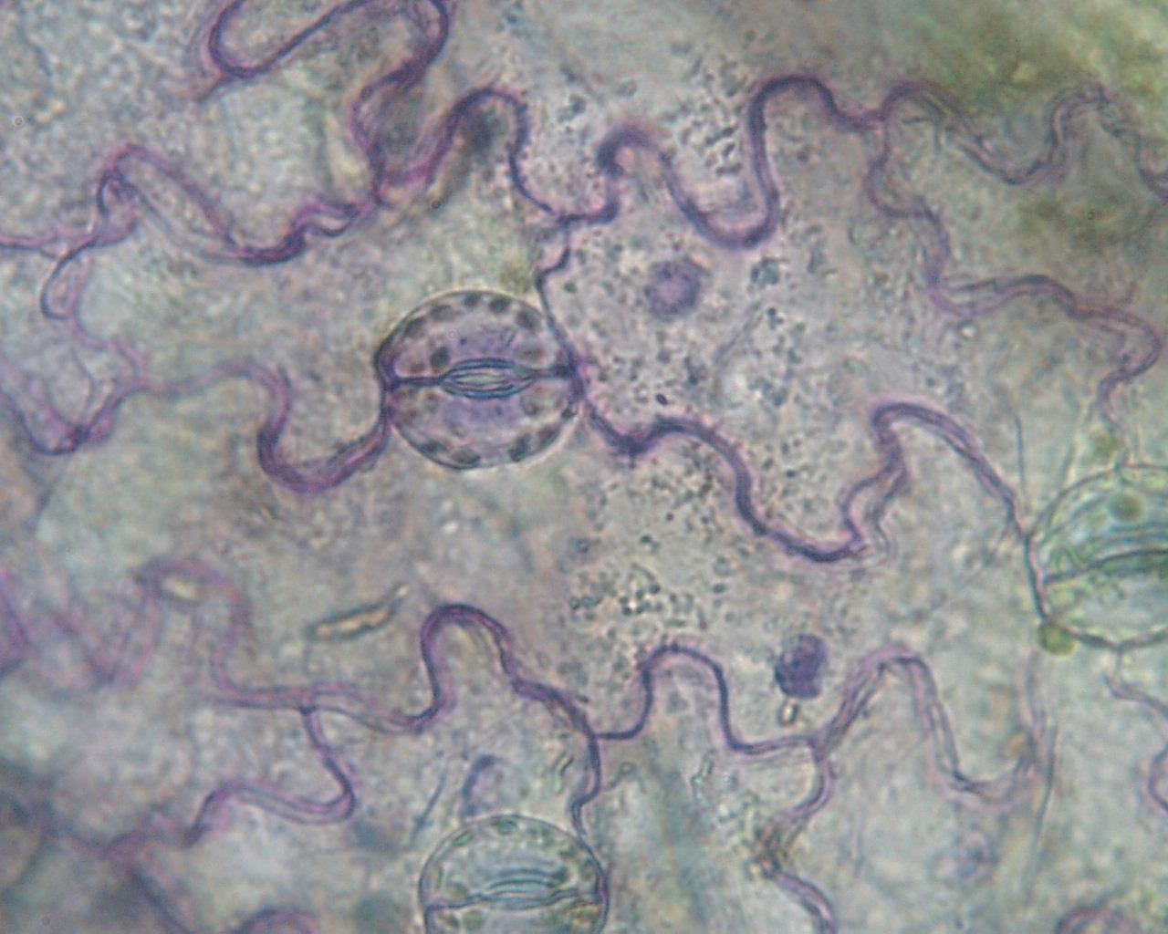
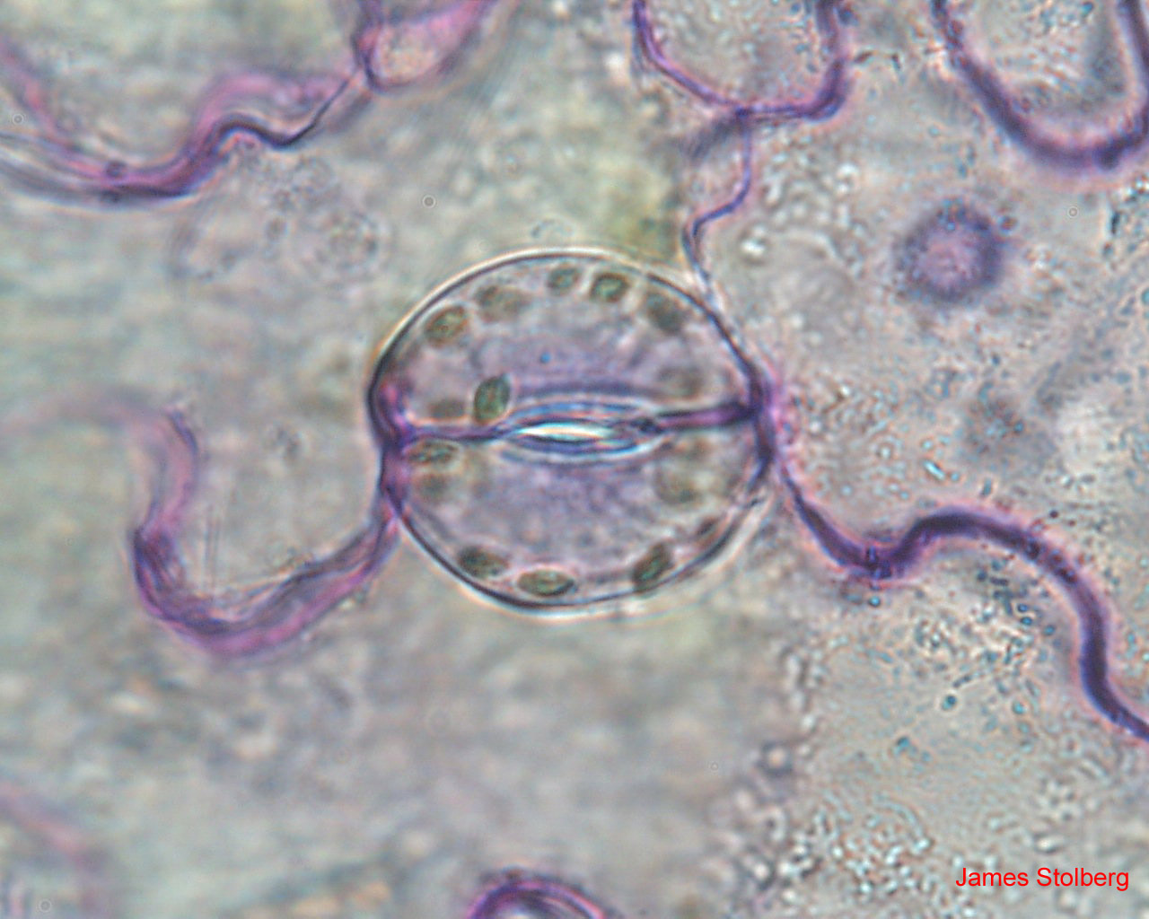
One enterprising group obtained some nice pictures from an epidermal
peel of a pea leaf that showed epidermal cells including guard cells. The
last image was collected from a 1000x oil immersion lens setup - look at
the chloroplasts!.
Lastly, one student noticed the presence of long trichomes coming out from the corn leaves. The image below is from a focus stacking of 8 images, each focusing one part of the trichome.