Lab 5 -- Monocot Roots
Here are the photos taken by students in class based on their hand
sections
from young corn roots. Labels have been added to some of
the images.
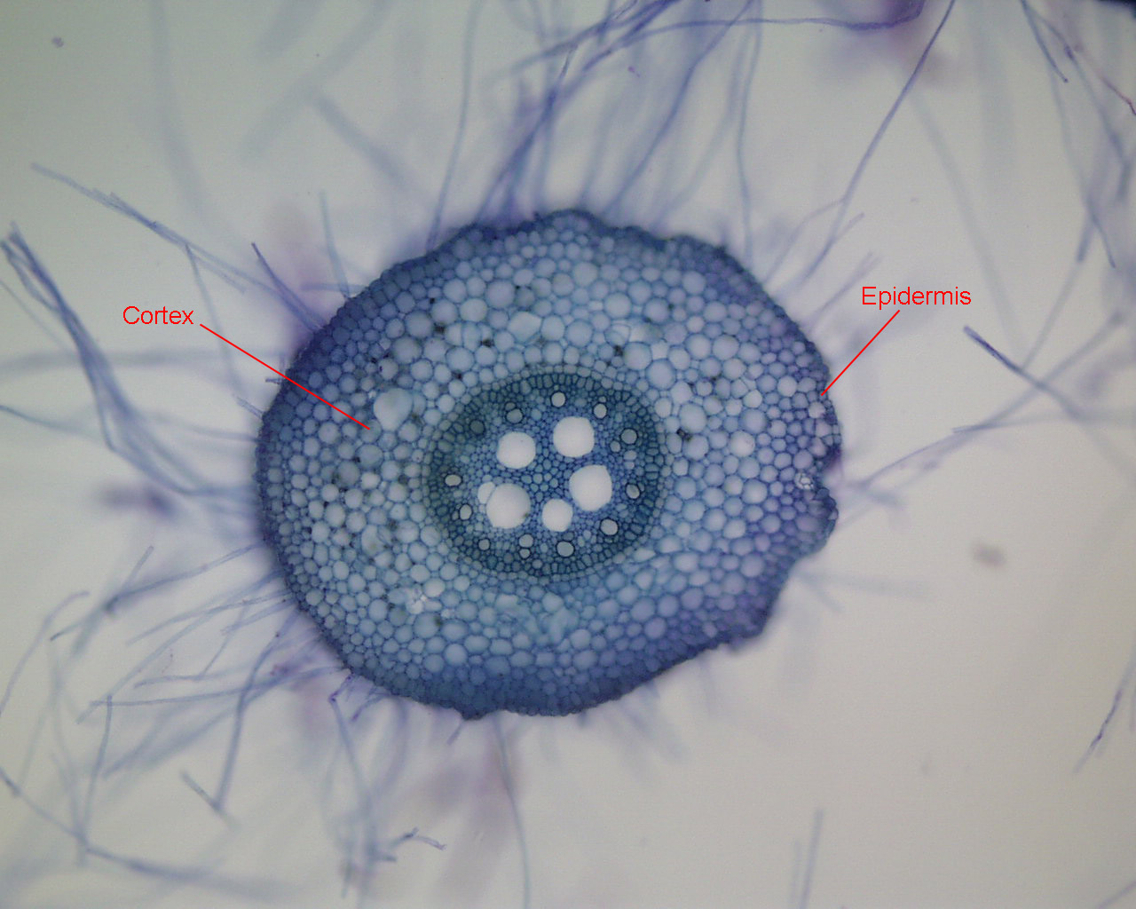
This first image shows the whole root. You can see how root hairs
can be very long and numerous.
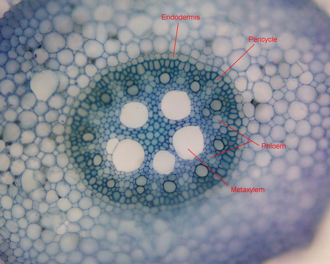
The next several images show closer views of the stele
Notice the exarch progression of protoxylem to metaxylem in this next image.
Here's a closer view of a newly forming root hair, where you can see
how it is an extension of an epidermal cell (no wall between the
epidermal cell and the root hair):

These next two images show the formation
(thickening) of the endodermis several centimeters back from the root
tip. Note the U-shaped appearance typical of many monocots.


This image shows the central region of the stele near the root tip, where the large metaxylem vessels are not yet mature and still have cytoplasm.

The next images show the production of branch roots